

On this page
A KEY QUESTION
Know what the record is for, how it will be taken and what checks are still needed at fitting.
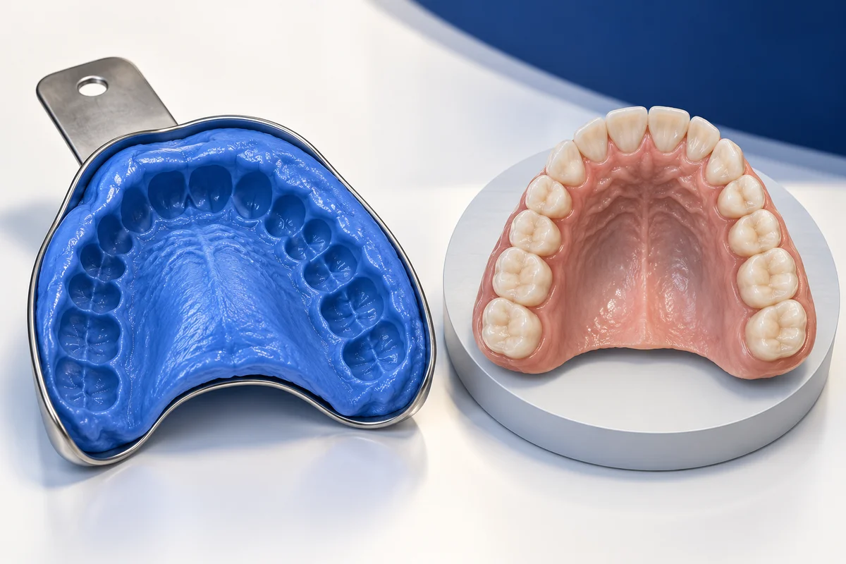
A conventional impression
A tray of soft material sits over the teeth until it sets, then is removed to provide a shape for a model. The upper jaw, lower jaw or both may be recorded. Describe any previous difficulty and find out what the sequence involves.
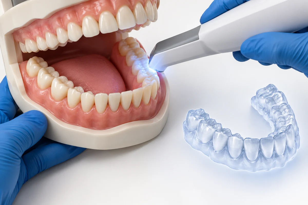
An intraoral scan
A wand collects surface images that a computer combines into a three-dimensional model. This can help discussions and laboratory work. The scanner still needs access around the teeth. Mention restricted opening or sore areas; digital recording cannot guarantee comfort or eliminate gagging.
Surface images and X-rays
A surface model shows the outside of teeth and gums. Dental X-rays and CBCT investigate different structures, including roots and bone. If the term “3D scan” is unclear, establish which equipment is meant and what question the clinician needs it to answer.
Planning, making and fitting
A preliminary record supports planning; a final scan or impression may be taken after preparation to make a restoration. A bite record relates the two jaws. Have any on-screen model identified as current teeth, prepared teeth or a proposed design.
Making the appointment manageable
Tell the team about gagging, anxiety, breathing difficulties or previous reactions to dental materials. Agree a stop signal before the tray or wand enters your mouth. Positioning, pauses or another recording method may help, depending on the clinical task.
The laboratory and next visit
The clinician checks whether the record contains enough information; repeats are sometimes needed. Confirm the laboratory and fitting arrangements, and allow time for clinical checks. When travelling for care, scanning speed alone should not determine your itinerary.
Read the related guidance
Keep the wider treatment plan in view while comparing these options.
Choose questions to ask.
Select your questions and copy the list, or open it as a WhatsApp draft. You can edit the message before sending.
Your consultation questions
Untick any questions you don’t need.
3 selected · No contact details needed.
Common questions
About this information
Published by Best Smile Turkey, a health-tourism intermediary. Your treating dentist is responsible for assessment, treatment and care.
Use this information to prepare questions for your dentist. An examination is needed to advise on treatment. Photographs, models and previews cannot establish suitability or predict results.
Source checking is not a clinical review. How we use sources and images, or how to report a correction.
Sources and further reading
- Cleveland Clinic: dental impressions Physical and digital records, preliminary and final impressions, bite registration and gagging concerns.
- UCLH: digital dentistry and intraoral scanning Surface models of teeth and gums, patient communication and laboratory workflow.
- FDA: dental cone beam computed tomography X-ray-based CBCT and its distinction from surface recording.
- NHS: dental treatments explained An overview of crowns, bridges, veneers, implants and other options.
- General Dental Council: going abroad for dental treatment Questions to ask about the treating provider, follow-up arrangements and concerns.
These resources offer general guidance; their publishers do not endorse Best Smile Turkey or the treating provider. Manufacturer links identify products or records, not independently compared outcomes. NHS funding information concerns UK care, not treatment costs in Turkey.
Ask about this information or report a correction