

On this page
A KEY QUESTION
Describe the swelling and its timing to a clinician; do not use a sharp object to try to remove a stone.
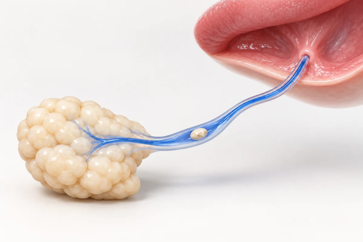
Where a stone can form.
Salivary glands make saliva, which reaches the mouth through small ducts. Many stones occur in the system beneath the tongue. A stone is not always visible.
A lump under the jaw or in the neck should not be labelled a stone without assessment. Other gland problems and unrelated conditions can produce swelling.
What to tell the clinician.
Note when the problem started, the side affected and whether swelling appears or intensifies during eating. Mention pain, a bad taste, discharge, fever or previous episodes. Tell the clinician if the swelling never fully settles.
You do not need to provoke the pain to demonstrate it. If an episode resolves before the appointment, your description and any photograph taken naturally may help, but cannot replace an examination.
How the next step is decided.
A GP or dentist can assess the problem and direct you to an appropriate service. Persistent or recurrent obstruction may need specialist investigation. Treatment depends on the position of a stone and what else the examination finds.
Some stones pass without a procedure; others need professional removal. Specialist services offer different approaches. Ask what the proposed treatment would do, whether further investigation is needed and how any infection would be addressed. Gland removal is not the inevitable outcome.
Do not dig for a stone.
Never cut, puncture or scrape the duct with a needle or another sharp object. Even a visible pale object may not be something you can safely remove. Pulling or forcing it can injure the area.
Follow advice suited to your health. If a clinician has restricted your fluid intake, do not replace that instruction with a general suggestion to drink more. A pharmacist or clinician can advise on comfort while you await assessment.
When to seek help.
Arrange assessment for troublesome swelling that persists or keeps returning. Pain with redness, pus, fever or feeling hot, cold or shivery may indicate infection and needs prompt medical advice.
Rapidly worsening mouth or neck swelling, or difficulty breathing, speaking or swallowing, needs emergency care. In the UK, call 999 or attend A&E; abroad, use the local emergency service. Do not wait to see whether a suspected stone passes.
Read the related guidance
Keep the wider treatment plan in view while comparing these options.
Questions for a clinical assessment
Seek the local care described above first. When you speak with the clinician, these questions may help you understand the plan.
- Who will carry out my treatment, and how can I check their qualifications and registration?
- Which options and alternatives should I discuss with the dentist?
- Who should I contact after returning home, and how will reviews be arranged?
Common questions
For future treatment planning, after appropriate clinical care: Compare starting prices in GBP, including zirconia crowns from £160. Your dentist confirms suitability and the full written quote.
About this information
Published by Best Smile Turkey, a health-tourism intermediary. Your treating dentist is responsible for assessment, treatment and care.
Use this information to prepare questions for your dentist. An examination is needed to advise on treatment. Photographs, models and previews cannot establish suitability or predict results.
Source checking is not a clinical review. How we use sources and images, or how to report a correction.
Sources and further reading
- NHS: salivary gland stones Symptoms, infection concerns, professional removal and the warning against sharp-tool self-removal.
- Guy’s and St Thomas’ NHS: salivary gland service Assessment of recurrent gland swelling and the range of specialist treatment approaches.
- NHS: dental abscess and emergency symptoms Emergency escalation for severe swelling affecting breathing, speaking or swallowing, without diagnosing its cause.
- NHS: dental treatments explained An overview of crowns, bridges, veneers, implants and other options.
- General Dental Council: going abroad for dental treatment Questions to ask about the treating provider, follow-up arrangements and concerns.
These resources offer general guidance; their publishers do not endorse Best Smile Turkey or the treating provider. Manufacturer links identify products or records, not independently compared outcomes. NHS funding information concerns UK care, not treatment costs in Turkey.
Ask about this information or report a correction